
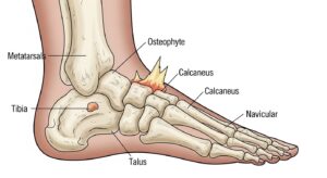
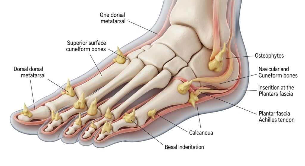
Bone spurs on the foot, particularly those forming on the heel bone known as heel spurs or calcaneal spurs, represent a common yet often misunderstood foot condition. These bony projections develop gradually as the body’s response to ongoing stress on the ligaments and tendons in the foot, most notably the plantar fascia—a thick band of tissue running along the bottom of the foot from the heel to the toes. While many individuals harbor these spurs without any discomfort, they frequently coexist with plantar fasciitis, the inflammation of that same plantar fascia, leading to significant heel pain that disrupts daily activities. Understanding this interplay is crucial for effective management, as the spur itself rarely causes pain; instead, it signals underlying tissue strain that requires targeted intervention.
This comprehensive guide walks you through every aspect of bone spurs on the foot, from their formation to proven relief strategies. Whether you’re experiencing sharp morning heel pain or seeking preventive measures, the steps outlined here draw from established medical approaches to empower you with actionable knowledge. By following a structured plan, most people achieve substantial improvement without invasive procedures, restoring mobility and comfort.
Understanding the Formation of Bone Spurs on the Foot
The process begins with repetitive microtrauma to the plantar fascia insertion point on the calcaneus, the large heel bone. Over time, the body’s repair mechanism deposits calcium, forming a bony outgrowth that protrudes from the underside of the heel. This adaptation aims to stabilize the stressed area but can exacerbate pressure if inflammation persists.
Primary Causes
Excessive strain from daily activities tops the list. Prolonged standing on hard surfaces, as seen in professions like teaching or nursing, repeatedly tugs at the fascia. High-impact exercises such as running on concrete amplify this, tearing the fascia’s covering and prompting spur growth.
Aging plays a pivotal role, with the plantar fascia losing elasticity after age 40, making it more susceptible to damage. Concurrently, the natural fat pad cushioning the heel thins, heightening impact forces.
Risk Factors in Detail
- Obesity: Excess body weight increases mechanical load on the feet, with each pound adding three times the pressure during walking. This chronic overload accelerates fascia degeneration, often leading to spurs within months of significant gain.
- Abnormal Foot Mechanics: High arches distribute weight unevenly, straining the heel, while flat feet cause overpronation, twisting the fascia excessively. Custom assessments reveal these imbalances early.
- Tight Calf Muscles: Shortened gastrocnemius and soleus limit ankle dorsiflexion, forcing the plantar fascia to bear more tension during gait. Daily stretching can mitigate this risk by up to 50 percent.
- Poor Footwear Choices: Flat shoes or worn-out soles lack arch support, allowing unchecked fascia stretch. Opting for cushioned, structured options prevents initial strain buildup.
- Repetitive Activities: Marathon training or dance routines without recovery periods create cumulative trauma. Incorporating rest days halves the incidence in active populations.
- Age-Related Changes: Post-40, reduced collagen in ligaments fosters rigidity, inviting spurs as a compensatory mechanism. Proactive mobility work counters this effectively.
- Previous Injuries: Healed ankle sprains alter gait patterns, overloading the heel long-term. Rehabilitation focusing on symmetry restores balance.
- Occupational Demands: Factory workers or servers endure constant hard-surface pounding, doubling spur likelihood. Scheduled breaks and supportive insoles provide immediate relief.
Recognizing these contributors allows for proactive adjustments, halting progression before symptoms emerge.
Identifying Symptoms of Bone Spurs on the Foot
The hallmark sign mimics plantar fasciitis: intense, stabbing pain at the heel’s bottom, peaking with the first steps after waking or prolonged sitting. This eases slightly after 10-15 minutes of movement but flares post-exercise or extended standing. Swelling may appear, accompanied by tenderness when pressing the arch-heel junction.
Advanced cases involve aching that radiates into the arch, limiting stride length and prompting limping. Nighttime throbbing disrupts sleep, while barefoot walking on tiles intensifies the burn. Differentiating from stress fractures requires professional evaluation, as both present similarly.
When Symptoms Signal Urgency
Persistent pain beyond two weeks, inability to bear weight, or numbness/tingling down the foot warrants immediate attention. These could indicate nerve compression or fascia rupture, complicating recovery.
Step-by-Step Guide to Accurate Diagnosis
Step 1: Self-Assessment at Home
Begin by noting pain patterns: Is it worse mornings? Does stretching calves alleviate it? Palpate the heel for a hard bump—though spurs are often deep. Track activity levels and shoe wear to share with your provider.
Step 2: Professional Physical Examination
Your doctor will inspect gait, foot posture, and arch height. Key tests include squeezing the forefoot to check medial compression pain and assessing ankle flexibility. Limited upward foot motion often confirms fascia tightness linked to spurs.
Step 3: Imaging Confirmation
X-rays visualize the spur as a hook-like projection from the calcaneus, ruling out fractures or arthritis. Views from side and angle pinpoint size and location. Rarely, ultrasound evaluates soft tissue inflammation, while MRI details fascia tears if surgery looms.
This methodical approach ensures precise identification, avoiding misdiagnosis of related issues like Achilles tendinopathy.
Comprehensive Treatment Strategies: From Home Remedies to Advanced Interventions
Treatment prioritizes symptom relief and fascia healing, as removing the spur alone seldom resolves pain. Over 90 percent succeed with conservative measures within 10 months, emphasizing patience and consistency.
Step 1: Immediate Home Care Protocol
Initiate the RICE method: Rest by avoiding aggravating activities, substituting swimming for running. Ice the heel 20 minutes thrice daily, rolling a frozen water bottle underfoot for massage. Compression via elastic wraps reduces swelling; Elevation post-activity minimizes fluid buildup.
Incorporate over-the-counter NSAIDs like ibuprofen to curb inflammation, following dosage guidelines to protect stomach lining.
Step 2: Targeted Stretching and Strengthening Routine
Dedicate 10 minutes twice daily to exercises proven to elongate the fascia:
- Calf Wall Stretch: Face a wall, place hands at shoulder height, extend one leg back with heel grounded, knee straight. Lean forward until stretch peaks in calf; hold 30 seconds, repeat 10 times per side. This lengthens the gastrocnemius, reducing heel pull by 40 percent over weeks.
- Plantar Fascia Towel Stretch: Seated, loop a towel around forefoot toes, gently pull toward shin while knee straight. Maintain 30 seconds, 10 reps. Ideal mornings, it counters overnight shortening for pain-free steps.
- Big Toe Extension: Sit, cross affected foot over knee, manually dorsiflex big toe upward 10 seconds, 15 reps. Targets intrinsic muscles, enhancing arch support and stability.
- Seated Calf Stretch: Loop towel under ball of foot, pull toes toward face with knee bent. Hold 30 seconds, 10 times. Isolates soleus muscle, vital for standing relief.
- Arch Roll: Stand, roll frozen golf ball under arch 5 minutes. Combines massage and cold therapy, dissolving adhesions daily.
- Ankle Circles: Seated, rotate ankle 20 times each direction. Boosts circulation, preventing stiffness buildup.
- Heel Drops: On step edge, lower heels slowly below toes, rise on balls. 15 reps. Eccentric loading strengthens tendons, accelerating recovery.
- Marble Pickup: Use toes to pick marbles into bowl, 20 times. Builds toe flexor power, redistributing foot pressure.
Step 3: Footwear and Orthotic Optimization
Transition to shoes with firm heel counters, ample cushioning, and moderate arch support—avoid flats or high heels. Silicone heel cups elevate the fat pad, offloading the spur site. Custom orthotics, molded from foam impressions, correct biomechanics permanently.
Step 4: Professional Therapies
Physical therapy amplifies home efforts with ultrasound, iontophoresis for deeper NSAID delivery, and myofascial release. Night splints maintain dorsiflexion overnight, slashing morning pain by 70 percent. For stubborn cases, extracorporeal shockwave therapy delivers acoustic waves to stimulate healing without downtime.
Step 5: Injection Therapies
Cortisone shots into the fascia provide rapid relief but limit to two annually to avoid rupture risk. Platelet-rich plasma, derived from your blood, promotes regeneration via growth factors, offering longer-term benefits.
Step 6: Surgical Considerations
Reserve for failures after 12 months: endoscopic plantar fascia release cuts tight bands, often excising large spurs. Gastrocnemius recession lengthens calves via small incision. Recovery spans 6-12 weeks with boot immobilization, yielding 85 percent success but potential nerve risks.
Monitor progress weekly; adjust as swelling subsides and strength returns.
Proven Prevention Strategies for Lifelong Foot Health
Forestall spurs by maintaining ideal weight—losing 10 pounds halves heel stress. Schedule calf stretches pre- and post-activity, fostering flexibility. Rotate shoes every 300-500 miles, inspecting for compression.
Low-impact cross-training preserves joints: cycle thrice weekly, interspersing walks on grass. Ergonomic mats under workstations absorb shocks for standers.
Annual podiatric checkups catch misalignments early, prescribing preventive insoles.
Conclusion
Bone spurs on the foot, though permanent, need not dictate your mobility. By addressing root causes like fascia strain through meticulous home care, targeted exercises, supportive gear, and timely professional input, over 90 percent reclaim pain-free steps. Embrace prevention as routine—weight management, smart footwear, and consistent stretching safeguard against recurrence. Consult specialists promptly for tailored plans, ensuring heels support your active life indefinitely. With diligence, heel health becomes a sustained reality, free from the shadow of spurs.